
From Philly and the Pa. suburbs to South Jersey and Delaware, what would you like WHYY News to cover? Let us know!
The CT machine stood ready like a giant white gleaming donut, but the patient wasn’t quite cooperating.
It was evening, and the third floor of the Pavilion at the Hospital of the University of Pennsylvania was mostly empty. Medical testing and imaging during regular business hours were done for the day.
But in this one exam room, half a dozen people crowded around the CT scanner bed, trying to position the patient just right.
“Can we move it?” asked Leening Liu, a PhD student at the University of Pennsylvania, after examining one angle. “Let’s go this way more.”
The patient on the bed wasn’t of flesh and bone, and there was no disease that the machine could scan for. Instead, this was a 19th-century double bass, the largest of the string instruments, on loan from the Philadelphia Orchestra.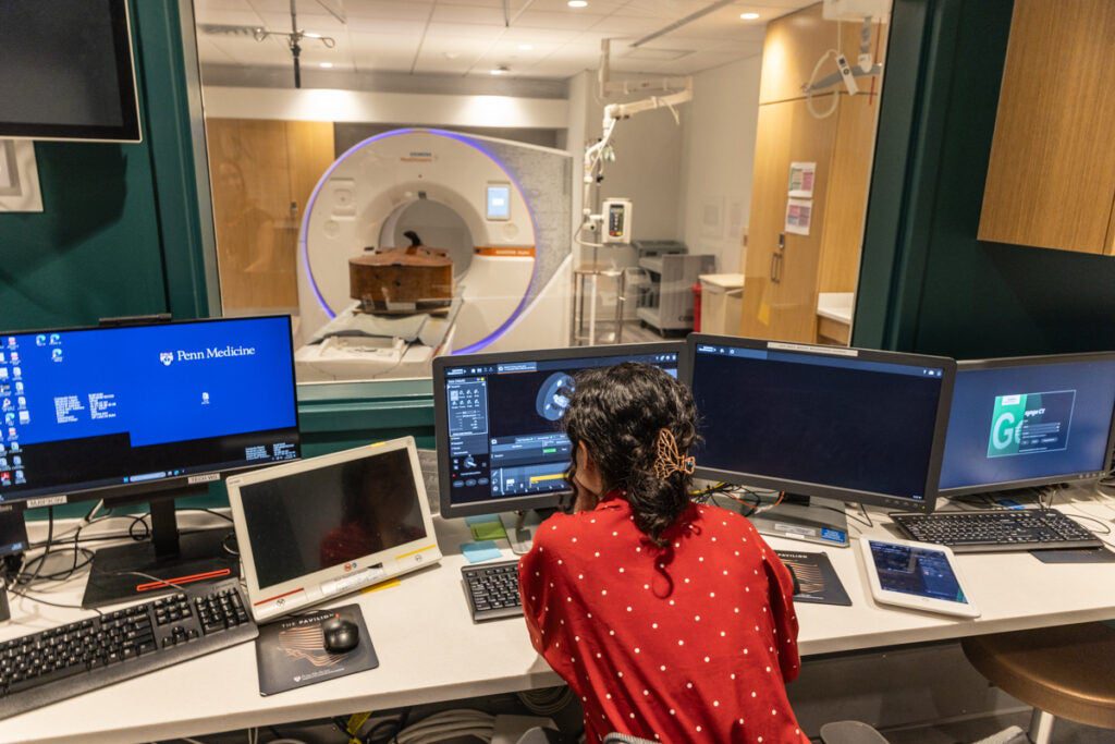 Researchers at the University of Pennsylvania are scanning historic instruments to help with their restoration. (Kimberly Paynter/WHYY)
“It’s much lighter than a comparable patient,” joked Peter Noël, an associate professor of radiology at Penn and director of CT research at the hospital.
CT scanners use some radiation to quickly produce detailed images of the inside of people’s bodies. They’re used to check for diseases like cancer, issues like blood clots, or injuries to the brain and internal organs.
They can also offer an inside view of inanimate objects and shed light on materials, production methods, age or the scars of time. Noël likes to use this medical technology for this kind of sleuthing research to unlock the mysteries of these objects. Scanning string instruments is his latest project.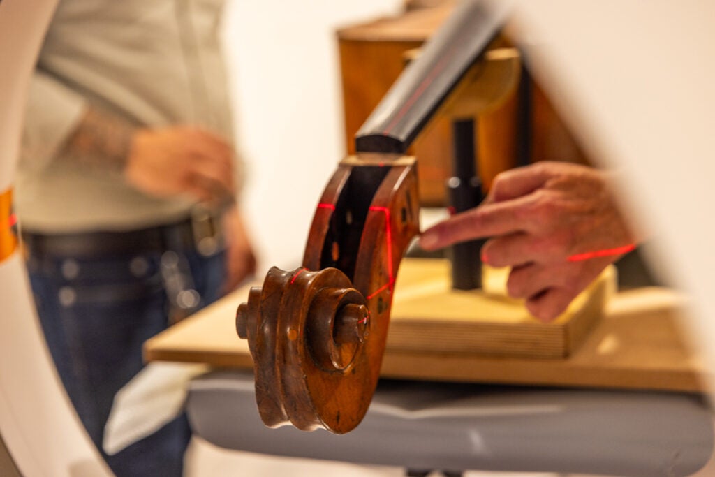 The scroll of the 19th century bass was adjusted multiple times to get the correct orientation for the CT scan. (Kimberly Paynter/WHYY)
The goal of imaging older double basses is to find out how to best preserve and restore these rare and priceless instruments, and how to make new ones more sustainably without sacrificing their unique low and deep “voices.”
“There are so many examples of where imaging can help, and I think that’s also where we can connect fantastically with the community, because these are everyday problems,” Noël said. “I mean, I don’t know if a bass is an everyday problem, but it’s a more positive thing and I think we can give back in this way.”
The double bass being scanned is a massive instrument over 5 feet tall. The four strings that typically run down the center had been removed for scanning. It’s considered a rare model and insured at over $200,000.
However, the bass’s owner, Mark Kindig, of Bethesda, Maryland, said it’s likely worth more in sale value, as that takes into account any previous famous players and owners, the quality and condition of the piece, its age, and the original luthier who crafted it in the first place. Peter Noël, PhD is the director of CT research at the Perelman School of Medicine in Philadelphia. (Kimberly Paynter/WHYY)
The double bass at the hospital was made an estimated 175 years ago by luthier Charles Theress.
The CT scan takes slices of images that can show the density of the wood. It can also reveal other characteristics, like how the wood has dried or warped over time, or what kind of repairs may have been made in the past.
“These scans, certainly in a restoration aspect, are very valuable because you can detect termite infestation, wood worms, damage that is subterranean, that you can’t typically see,” said Zachary Martin, a luthier, bass restorer and maker from Providence, Rhode Island.
Source link : http://www.bing.com/news/apiclick.aspx?ref=FexRss&aid=&tid=66f2a6184a8244c99ff3782a838aabe7&url=https%3A%2F%2Fwhyy.org%2Farticles%2Fmedical-imaging-ct-scan-philadelphia-orchestra-environment%2F&c=1523484223419412954&mkt=en-us
Author :
Publish date : 2024-09-24 00:17:00
Copyright for syndicated content belongs to the linked Source.